
Written by Shivani Verma, IAFR Member
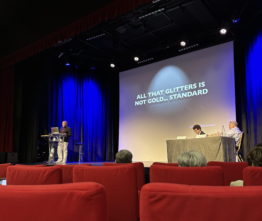
Forensic imaging is a powerful modern innovation that aims to detect and document findings in forensic investigations – primarily for anthropology, archaeology, post-mortem and medicolegal purposes. The 2023 ISFRI conference hosted in Toulouse, France was a show stopping exhibition that presented new findings, and interesting cases supported by forensic imaging. The conference served as the 12th Annual Meeting of the International Society of Forensic Radiology and Imaging and the 18th Anniversary Meeting of the International Association of Forensic Radiographers.
Presenters and attendees from 25 different countries were invited to the ISFRI congress. Some of the presentation categories included Trauma & Post Mortem CT (PMCT), Anthropology and Fossil Imaging, Forensic Ballistic Imaging and Forensic Paediatric Imaging.
The majority of all radiological techniques currently used in forensic medicine are derived from clinical practice, including conventional radiography, MRI, CT, and CT angiography.
Conventional x-ray is also much quicker to acquire than other modalities and is good in identifying age estimation variables. Unconventional x-rays include LODOX scanners which produce low dose x-ray, originating from South Africa, where a patient can be x-rayed head to toe in less than 1 minute. Efithmia Letiz led a brilliant research study on the comparison of LODOX and conventional radiography.
PMCT primarily focuses on traumatic events such as traffic accidents, falls from heights, blunt force violence and gunshot incidents. PMCT is also an important tool in cases of infant death, where it is useful for estimating age, especially for bodies lacking an identity. Multi-phase post mortem CT-angiography (MPMCTA) is the most used and researched technique for post mortem whole body perfusion. This minimally invasive technique incorporates a specific perfusion device pump an injection into the venous and arterial
systems.
MRI is commonly used for the diagnosis of natural deaths and for the assessment of traumatic soft tissue injuries. It has been used successfully in several facilities in the investigation of fatal neonate and infant malformations.
Usefulness of Virtual Reassembly of skull or spine in case of fragmentation due to high energy trauma or bone separation through postmortem changes by Haruki Fukuda
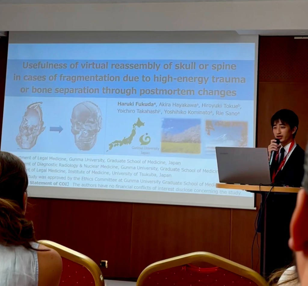
High energy trauma is commonly associated with road traffic accidents, collisions, crushing accidents and falls typically result in complex injury patterns. Haruki Fukuda presented on the ‘Usefulness of Virtual Reassembly of skull or spine in case of fragmentation due to high energy trauma or bone separation through postmortem changes’. He is a forensic pathologist from Japan whose study aimed to identify whether virtual reassembly of the skull or spine is more effective than adhesive reconstruction.
He showcased three cases, all from high-impact scenarios, to assess the visualisation of the fractures using these two reconstruction methods. High energy trauma makes it difficult to analyse microscopic changes due to the multitude of bony fragments. In all three cases he concluded that virtual reconstruction is more useful in visualising the number and extent of fractures, if the correct software is used. These reconstructions can also be made in a more timely manner than adhesive reconstruction.
The complementary roles of whole-body post mortem computed tomography and conventional skeletal survey by Selena de Vries et al.
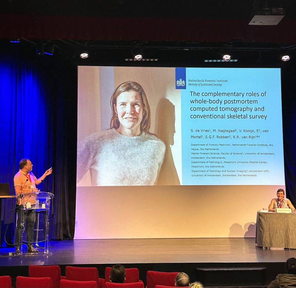
Another study pertinent to forensic imaging is one conducted by Selena de Vries examined the use of whole body PMCT scans alongside conventional skeletal surveys for the detection of injuries prior to forensic autopsy in children.
This was a retrospective study with data acquired from 116 cases between 2008 and 2021. Fractures were seen in 44% of the cases examined, with a total of 206 fractures identified. When compared to conventional skeletal surveys, PMCT was able to detect more fractures particularly rib fractures, with 90% of rib fractures missed on x-ray.
However, conventional x-ray performed better when used for examination of extremities. An example of why this may have been the case can be seen when x-raying the hand. The hands of deceased children will naturally form a clenched fist post mortem. When taking an x-ray, positioning techniques can be used to stretch the hand out. While in CT, the hand will likely remain clenched during imaging.
Detection of iodine using spectral imaging on PMCT by Chris O’Donnell and Catherine Vincent
An incredible presentation given by Chris O’Donnell, who is a Radiologist at the Victorian Institute of Forensic Medicine, discussed how spectral imaging/dual energy CT can detect different material compositions within deceased bodies.
This technique can be used to examine the presence of iodine contrast in the body and determine whether contrast seen on imaging was injected before or after death. Which is important, as many patients tend to have contrast CT scans prior to their deaths, and in some cases there may not be not enough time for renal excretion of contrast to occur between the time of injection and patient death. This ultimately results in leftover iodine contrast within the body mimicking other pathology such as diffuse hypoxic ischemic injury on post mortem CT imaging.
Forensic Imaging is a revolutionising and ever-evolving field of Medical Imaging. In Australia we don’t have the forensics rates such as the UK or the Americas so an increased awareness of how imaging can be utilised in other ways is pertinent to essentially opening up training and further research opportunities.
A huge thank you to the IAFR for giving me the opportunity to attend this conference!